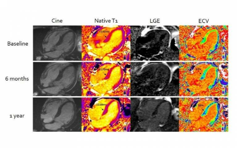
MRI with CMR (T1) & ECV mapping - Cardiac amyloidosis improvement demonstrated by Cardiovascular Magnetic Resonance (CMR) Extracellular Volume Mapping (ECV) for amyloid. Credit UCL.
MRI with CMR (T1) & ECV mapping - Cardiac amyloidosis improvement demonstrated by Cardiovascular Magnetic Resonance (CMR) Extracellular Volume Mapping (ECV) for amyloid. Credit UCL. An advanced form of cardiac MRI, developed by academics at UCL in collaboration with the Royal Free Hospital, has for the first-time enabled clinicians to measure the effectiveness of chemotherapy in patients with the life-limiting condition 'stiff heart syndrome'. Researchers say the breakthrough, published in the European Heart Journal , means doctors will now be able to better guide treatment strategies and, by doing so, improve patients' prognosis. Light-chain cardiac amyloidosis (stiff heart syndrome) occurs when plaques of protein called amyloid build up in heart muscle, affecting its ability to pump blood, and without treatment can rapidly lead to heart failure and death. However, assessing the condition has been difficult, as while clinicians can detect the presence of amyloid in the heart, there has been no safe test to measure the amount. This has also meant there has been no way of measuring the therapeutic effect of chemotherapy - the normal first line treatment.
TO READ THIS ARTICLE, CREATE YOUR ACCOUNT
And extend your reading, free of charge and with no commitment.