New imaging technique could lead to better bio-implants for patients
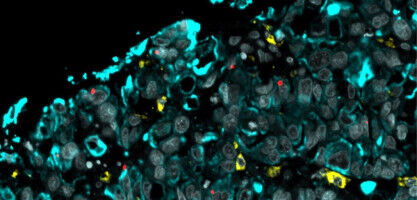
University of Birmingham scientists have developed a new microscopic imaging approach to take a closer look at 3D-printing for developing future patient implants, as well as improved disease modelling and drug screening. Additive manufacturing (3D printing) platforms create bioprinted structures by moving a special bioink, containing cells, biomolecules and materials, through a narrow tube, but the process can result in cells becoming damaged as they pass through the tiny tube. Using a microscopy technique that shines a blade of light in the material flowing inside the narrow tube, the researchers have been able to examine and reveal important information about how cell damage can occur during the bioprinting process. The imaging process could give better understanding of bioink flow dynamics and cell movement - enabling investigation of complex capillary designs and process to improve 3D bioprinting and printed constructs. Publishing their findings in Bioprinting today, the scientists reveal that their imaging technique illuminates a range of hydrogel-cell behaviour and damage patterns depending on the extrusion speed and properties of the tube used during printing. Report co-author Dr. Gowsihan Poologasundarampillai , Fellow in Biomaterials and Bioimaging at the University of Birmingham's School of Dentistry, commented: "Additive manufacturing platforms are transforming research and manufacturing worldwide. We used light sheet fluorescence microscopy (LSFM) to mimic the portion of the extrusion bioprinting process in which cells are most likely to be damaged.