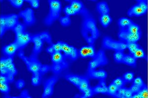
Individual proteins are clusters at the surface of tat the surface of the cell
24 Jul 2013 Scientists from The University of Manchester have revealed new images which provide the clearest picture yet of how white blood immune cells attack viral infections and tumours. They show how the cells, which are responsible for fighting infections and cancer in the human body, change the organisation of their surface molecules, when activated by a type of protein found on viral-infected or tumour cells. Professor Daniel Davis, who has been leading the investigation into the immune cells, known as natural killers, said the work could provide important clues for tackling disease. The research reveals the proteins at the surface of immune cells are not evenly spaced but grouped in clusters - a bit like stars bunched together in galaxies. Professor Davis, Director of Research at the Manchester Collaborative Centre for Inflammation Research (MCCIR), a partnership between the University and two pharmaceutical companies GlaxoSmithKline and Astra Zeneca, said: "This is the first time scientists have looked at how these immune cells work at such a high resolution. The surprising thing was that these new pictures revealed that immune cell surfaces alter at this scale - the nano scale - which could perhaps change their ability to be activated in a subsequent encounter with a diseased cell. "We have shown that immune cells are not evenly distributed as once thought, but instead they are grouped in very small clumps - a bit like if you were an astronomer looking at clusters of stars in the Universe and you would notice that they were grouped in clusters.
TO READ THIS ARTICLE, CREATE YOUR ACCOUNT
And extend your reading, free of charge and with no commitment.